Canine Splenic Masses and Internal Bleeding: From Emergency Triage to Tissue Diagnosis
Summary
An owner-focused review of splenic-mass emergencies, stabilization, imaging limits, surgical diagnosis, and decisions under uncertainty.
Article
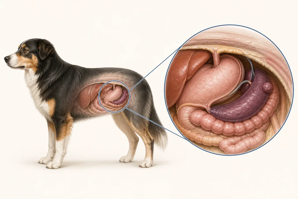
High-urgency evidence review. A splenic mass may be discovered incidentally or after internal bleeding causes sudden weakness or collapse. Benign and malignant splenic lesions can present similarly, and imaging generally cannot provide the final tumor diagnosis. The immediate task in an unstable dog is stabilization; the definitive answer commonly depends on microscopic examination of removed tissue.
Emergency presentation
Possible signs include weakness, fainting or collapse, pale gums, a distended abdomen, rapid breathing or heart rate, reduced appetite, and waxing-and-waning lethargy. A bleeding mass can temporarily clot, so a dog may appear to improve and then deteriorate again. Apparent recovery is not proof that the danger has passed. Collapse, pale or white gums, labored breathing, marked abdominal enlargement, or severe weakness warrants emergency veterinary care immediately.
Initial clinical priorities
The emergency team evaluates circulation, breathing, blood loss, pain, and concurrent disease. Testing may include blood counts and chemistry, clotting assessment, abdominal fluid evaluation, radiographs, ultrasound, and cardiac monitoring. Intravenous fluids, blood products, oxygen, or intensive monitoring may be necessary before anesthesia. The appropriate sequence depends on stability.
Ultrasound can identify a splenic mass and free abdominal fluid and can examine other organs. Chest imaging or echocardiography may be recommended in some cases to look for disease that changes prognosis or anesthetic planning. These studies estimate likelihood and extent; they do not reliably distinguish every hematoma, benign nodule, hemangioma, hemangiosarcoma, or other lesion.
Why the diagnosis often follows surgery
According to the American College of Veterinary Surgeons, splenectomy is the primary treatment for many splenic masses and can be both diagnostic and therapeutic. Sampling a vascular splenic lesion before surgery may not be the safest or most informative choice in every patient. After removal, the mass should be submitted for histopathology. A presumptive label based on age, breed, rupture, or ultrasound appearance remains presumptive until tissue is examined.
Decision-making under time pressure
Owners may need to decide about surgery before knowing whether a lesion is benign or malignant. Ask the clinician to separate facts from probabilities and to explain:
- the dog's current stability and immediate risk of further bleeding;
- what imaging shows about the spleen, abdomen, chest, and heart;
- anesthetic and surgical risks, including hemorrhage and arrhythmia;
- whether surgery could be therapeutic if the lesion is benign;
- likely next steps for each major pathology outcome;
- the comfort-focused alternative if surgery is not consistent with the dog's condition or family goals.
After surgery
Monitoring may include heart rhythm, red-cell measures, pain, incision, appetite, vomiting, and signs of continued bleeding or other complications. At home, follow discharge instructions for activity restriction, collar use, medication, and incision checks. Do not infer the diagnosis from how quickly the dog feels better. Schedule the pathology-results discussion and request the complete report.
Interpreting prognosis
Outcome differs greatly between benign lesions and malignant tumors and also varies with stage, rupture, metastasis, other disease, and treatment choices. Population percentages can support a conversation but cannot tell an individual family what the pathology will show. Avoid products or testimonials claiming to identify or treat a splenic tumor without tissue diagnosis and veterinary planning.
Medical disclaimer: This article is educational and is not a diagnosis, triage service, or surgical recommendation. Suspected internal bleeding is life-threatening. Seek emergency veterinary care immediately for collapse, pale gums, rapid or difficult breathing, sudden abdominal distension, severe weakness, or rapid deterioration.