Canine Liver Masses: Pattern, Tissue Identity, and Hemorrhage-Aware Planning
Summary
A focal liver mass, multiple nodules, and diffuse change have different differentials, yet imaging pattern alone cannot reliably supply tumor type or predict every bleeding risk.
Article
Evidence-informed clinical review. The liver can contain a primary tumor, metastasis, lymphoma, benign nodules, cystic lesions, inflammatory change, or several processes at once. Ultrasound and CT reveal distribution and surgical anatomy, but their appearance is not a pathology report. The decision to sample, remove, monitor, or stabilize must reflect both diagnostic value and hemorrhage risk.
The clinical question
The useful question is what information is needed next: proof of tissue type, mapping for possible lobectomy, staging for disease elsewhere, or explanation of systemic illness. The answer changes when a dog is stable with an incidental lesion versus anemic, painful, jaundiced, coagulopathic, or showing evidence of abdominal bleeding.
What current evidence can establish
Laboratory testing describes liver function and systemic consequences but does not name most masses. Imaging can distinguish massive, nodular, and diffuse patterns and assess vessels, bile ducts, other organs, and abdominal fluid. Cytology may answer selected questions; core biopsy or surgical histopathology may be needed for architecture and classification.
A decision-focused pathway
Review trend data, not a single enzyme value. Confirm platelet and coagulation context, assess abdominal fluid when indicated, and stage according to the leading differential and intended intervention. Before percutaneous sampling, define the route, the expected diagnostic yield, what will happen if the result is nondiagnostic, and how bleeding will be detected and managed.
If surgery is discussed, the surgeon should evaluate lobe location, vascular and biliary relationships, other hepatic lesions, extrahepatic disease, and anticipated residual function. The pathology submission should preserve orientation and include clinically relevant history. Postoperative decisions depend on histology, margins where meaningful, spread, recovery, and family goals.
Evidence gaps and interpretation
Published prognoses vary sharply among hepatocellular, biliary, vascular, neuroendocrine, mesenchymal, and metastatic tumors. A favorable report for one resectable subtype cannot be generalized to every hepatic mass. Conversely, an alarming image does not remove the need to identify what disease the image actually represents.
Safety, monitoring, and escalation
Collapse, pale gums, abdominal distension, severe weakness, jaundice with illness, or uncontrolled vomiting requires prompt assessment. Liver sampling and surgery can cause hemorrhage, bile leakage, infection, pain, or organ dysfunction. Families need explicit emergency, medication, feeding, incision, and recheck instructions.
Distribution and bleeding risk guide the route to diagnosis
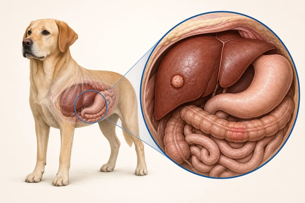
A single large hepatic mass, multiple nodules, and diffuse parenchymal change create different diagnostic and procedural questions. Imaging should describe the affected lobes, relationship to major vessels and the gallbladder, abdominal fluid, evidence of rupture, and lesions in other organs. These patterns can prioritize possibilities, but overlap is substantial and pattern recognition alone cannot supply a final tumor type.
Before tissue collection, the team can connect hemodynamic stability, platelet and coagulation information, lesion accessibility, and the amount of tissue required to the proposed method. A small aspirate, core sample, or surgical specimen does not carry the same information or risk. If hemorrhage is suspected, immediate stabilization and a bleeding-aware plan take priority over completing an idealized staging sequence. The record should state which unanswered question the chosen specimen is intended to resolve.
Questions for the veterinary team
- Is the liver change focal, multifocal, or diffuse?
- What tissue result would change management?
- How has bleeding risk been assessed?
- Does imaging support a safe local procedure?
- Which pathology and staging findings determine follow-up?
Medical disclaimer: This article is educational and cannot diagnose, treat, or determine prognosis for an individual dog. It does not replace an examination or an individualized plan from a licensed veterinarian.