Canine Mammary Masses: Mapping Every Nodule Before Selecting Surgery
Summary
A structured approach to documenting multiple mammary nodules, establishing histology, staging selectively, and matching surgery to anatomy rather than assumptions.
Article
Educational medical disclaimer: This article is for general education only and cannot determine whether a mammary nodule is benign or malignant or choose an operation for an individual dog. Any new or changing mammary mass should be examined by a veterinarian.
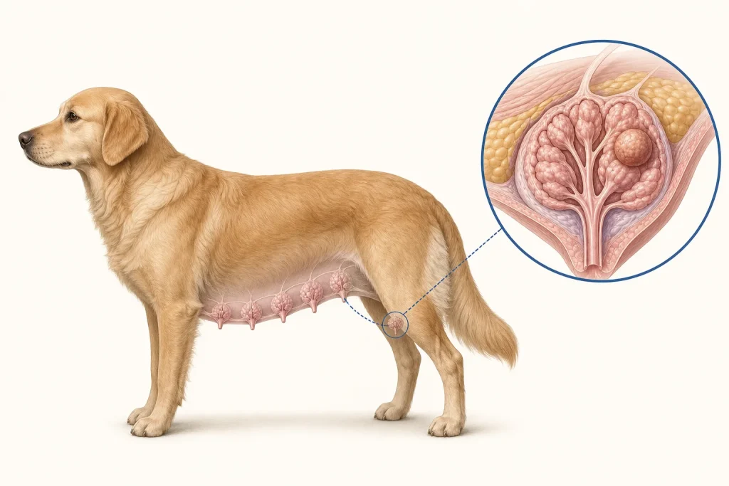
A dog has two mammary chains and may develop more than one nodule at the same time. Those nodules can differ in size and even in histologic type. Treating the case as one generic breast lump can lose information that matters for sampling, surgery, pathology labeling, and follow-up. The first practical task is to create a map.
Map the entire field
Each gland and nodule should be identified by side and location, measured in three dimensions, and described for mobility, attachment, surface change, ulceration, and discomfort. The clinician also examines regional lymph nodes and the rest of the dog. A dated diagram and photographs can prevent confusion when several lesions are close together. Owners should report growth history, reproductive status, prior mammary surgery, medications, and changes in appetite, weight, breathing, or activity.
Sampling can answer different questions
Fine-needle aspiration may help distinguish a mammary-origin lesion from inflammation or another mass, and suspicious lymph nodes may be sampled. However, cytology may not always separate benign from malignant mammary tumors because architecture and invasion matter. A 2024 study evaluating fine-needle and core-needle approaches illustrates that test performance depends on the sample and question; it does not make one technique universally definitive.
Histopathology of representative or removed tissue is central. It can identify tumor type and evaluate features associated with behavior. A study of 350 canine mammary tumors demonstrates the histologic diversity encountered in practice. When multiple nodules are removed, each should be clearly labeled by site rather than pooled without orientation. Otherwise, a clinically important result may be impossible to match to the correct location.
Stage according to established risk
The American College of Veterinary Surgeons describes evaluation that may include laboratory testing, thoracic imaging, and lymph-node assessment. The exact workup should reflect clinical findings, pathology, planned intervention, and family goals. Node size alone is imperfect: reactive nodes can enlarge, and metastatic cells can occur in nodes that feel normal. Sampling is considered when the answer could change the plan. Negative imaging means no spread was detected by those tests; it cannot exclude microscopic disease.
Surgery is tailored, not automatically maximal
Operations may range from removal of an individual mass to removal of one or more glands or a chain, depending on number, distribution, size, fixation, blood supply, prior surgery, and surgeon judgment. Wider surgery can create more wound tension and recovery burden without necessarily adding the same value in every case. Conversely, removing only the visible surface nodule without planning can compromise margins or miss a separate lesion. Owners should ask how the proposed field was chosen and how every specimen will be oriented for pathology.
Additional treatment or surveillance depends on final histology, grade where relevant, margins, stage, healing, and the dog's health. Statements about hormones, chemotherapy, or other therapies need to be tied to the exact diagnosis and evidence rather than applied to all mammary tumors.
Questions before consent
- How many nodules are present, and are they all included on the map?
- Which lesion or node should be sampled before surgery?
- What staging result would alter the operation or another treatment?
- Why is this specific surgical extent recommended?
- How will specimens be labeled so pathology findings return to the right site?
- What wound, mobility, and pain-support plan should be prepared at home?
- How will remaining glands and regional nodes be monitored?
Urgent veterinary contact is warranted for uncontrolled bleeding, a rapidly opening or infected wound, marked weakness, difficult breathing, or severe pain. Otherwise, owners can help by avoiding repeated squeezing, keeping a clean measurement log, and bringing all prior pathology reports to the next visit. A good mammary-tumor plan is not built from nodule size alone; it links the whole-chain map, tissue diagnosis, selective staging, and realistic recovery goals.