A Dog With New Seizures and a Brain Mass: Separating Imaging Suspicion From Tissue Diagnosis
Summary
An evidence-aware framework for discussing MRI findings, presumptive diagnoses, sampling limits, and goal-based care after a canine brain mass is detected.
Article
Educational medical disclaimer: This article cannot diagnose a brain disorder or recommend treatment for an individual dog. A first seizure, repeated seizure, prolonged seizure, collapse, severe disorientation, or rapidly worsening neurologic function needs prompt veterinary care.
A brain mass discovered after seizures or behavior change can create pressure to name the disease immediately. Magnetic resonance imaging is powerful, but the responsible interpretation often remains a presumptive diagnosis unless tissue has been examined. The central challenge is to use imaging, neurologic findings, and the dog's overall condition to make proportionate decisions while being honest about what remains unknown.
Stabilization comes before classification
Immediate priorities may include controlling seizures, managing dangerous pressure effects, protecting the airway, and checking for metabolic problems that can also cause neurologic signs. Owners should record the start and stop time of an event, video it only if safe, move hazards away, and follow the emergency instructions supplied by their clinic. They should not put hands in the dog's mouth or improvise extra medication.
MRI describes location and behavior
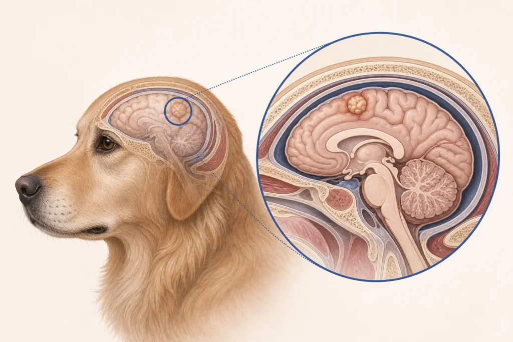
MRI can characterize where a lesion sits, its relationship to the meninges and ventricles, surrounding swelling, contrast enhancement, mass effect, and evidence of hemorrhage or other abnormalities. Those features help construct a differential list and may support a presumptive tumor category. A 2024 scoping review found substantial variation in MRI methods and reporting across canine primary brain-tumor studies. This reinforces a practical point: imaging patterns overlap, and the exact tumor type or grade may not be reliably determined from appearance alone.
The radiology report should be read as calibrated evidence. Terms such as most likely, compatible with, and cannot exclude carry different levels of certainty. Owners can ask the neurologist or oncologist which features support the leading diagnosis, what important alternatives remain, and whether additional imaging elsewhere in the body would alter the differential.
When tissue is considered
Histopathology can provide a more definitive tumor identity and, when applicable, grade. Biopsy or surgery may be reasonable when the lesion is accessible, the answer could change care, and procedural risk is acceptable. In other cases, location, patient health, cost, or family goals may make a presumptive approach more appropriate. Choosing not to biopsy does not transform an imaging impression into certainty; the medical record should preserve that distinction.
Treatment options answer different goals
Surgery may obtain tissue, reduce mass effect, and provide local control in selected lesions. Radiation therapy can address lesions that are not readily removed and may be used alone or after surgery. Medication may control seizures, inflammation, pain, nausea, or other signs; specific drug selection and tapering require veterinary direction. Systemic therapy has a role for some tumor types or situations. Comfort-focused care is also active care when burdens of diagnosis or treatment outweigh likely benefit.
A multicenter retrospective study of canine glioma treatments found outcome differences among treatment groups, but retrospective comparisons are vulnerable to selection bias and differences in case characteristics. Such data help frame questions; they do not predict an individual result or prove that the same option is appropriate for every brain mass.
Build a home neurologic baseline
- Keep a seizure log with duration, recovery time, possible clusters, and medication timing.
- Track walking, circling, vision-related collisions, sleep, appetite, toileting, and interaction.
- Ask for written thresholds for an emergency visit and for use of any prescribed rescue medication.
- Use gates, rugs, and supervised stairs to reduce injury without isolating the dog.
- Schedule reassessment based on clinical risk, not only when a scan is due.
Decision questions
What is known versus presumed? Could tissue change the treatment? Is the primary objective seizure control, longer local control, tissue diagnosis, preserved daily function, or some combination? What anesthetic and neurologic risks matter? How will benefit be judged, and what is the backup plan after progression or unacceptable effects?
These questions turn a frightening image into a monitored care plan. They also give families permission to revisit choices as function, response, logistics, and goals change.