A Cranial Mediastinal Mass in a Dog: Thymoma, Lymphoma, and Myasthenia Risk
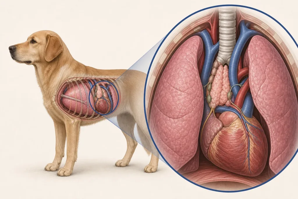
Summary
A mass in the front of the chest may compress airways or vessels and may be associated with neuromuscular disease; imaging location is a starting point, not a final diagnosis.
Article
Evidence-informed clinical review. Thymoma and lymphoma are important differentials for a cranial mediastinal mass, but other lesions can occur. Thymoma may be associated with acquired myasthenia gravis and megaesophagus, which can materially change anesthesia, aspiration, feeding, and surgical risk even when overt weakness is not the presenting complaint.
The clinical question
The team must determine respiratory stability, identify pleural fluid or compression, distinguish the leading tissue possibilities, and look for regurgitation, swallowing change, exercise weakness, or aspiration concern. The plan should answer whether sampling is safe and whether surgical anatomy and neuromuscular status support a local procedure.
What current evidence can establish
Thoracic radiographs identify the mediastinal pattern; CT can map extent, vessels, airways, lungs, nodes, and surgical relationships. Cytology or histopathology helps separate epithelial thymic tumor from lymphoma and other disease. Acetylcholine-receptor antibody testing and neuromuscular evaluation are considered when clinical suspicion for myasthenia exists.
A decision-focused pathway
Clarify whether the dog coughs, regurgitates, changes bark, fatigues, or struggles to swallow, because families may use vomiting to describe passive regurgitation. Assess oxygenation and pleural fluid before sedation. Plan fresh and fixed samples when lymphoma testing may require flow cytometry as well as cytology or tissue architecture.
For a potentially resectable thymoma, surgery planning must account for invasion, adjacent vessels, ventilation, and postoperative aspiration monitoring. Lymphoma generally shifts the discussion toward systemic therapy. When neither route offers an acceptable burden, radiation or symptom-focused care may be considered with a clearly defined goal.
Evidence gaps and interpretation
Older case series and small cohorts cannot predict an individual course, and the frequency of associated syndromes varies by population and definitions. A negative antibody test may not settle every neuromuscular question, and imaging cannot always distinguish thymoma from lymphoma. Treatment selection should reflect diagnostic confidence.
Safety, monitoring, and escalation
Breathing distress, blue or gray mucous membranes, collapse, repeated regurgitation, aspiration signs, or profound weakness needs urgent care. Fasting and medication instructions must be individualized because megaesophagus and respiratory compromise alter routine anesthesia preparation. Food or water should not be forced into a weak dog.
Do not let the chest mass hide a neuromuscular complication
A cranial mediastinal mass creates a focused differential, but imaging appearance alone may not reliably separate thymic, lymphoid, and other lesions. The initial record should pair respiratory signs and thoracic findings with lymph-node distribution, blood-cell information, and the least risky route to diagnostic tissue. Pleural fluid, vessel compression, or airway compromise can change the order in which those steps are taken.
Thymoma is also associated with acquired myasthenia gravis in some dogs, so regurgitation, swallowing difficulty, exercise-related weakness, and aspiration risk deserve deliberate review rather than being treated as unrelated complaints. Those signs can affect anesthesia and postoperative planning even before tumor treatment is selected. A negative symptom history is useful baseline information, while compatible signs call for targeted veterinary assessment rather than assumptions based only on the mass.
Questions for the veterinary team
- What is the dog’s respiratory and aspiration risk today?
- Does the pattern favor thymoma, lymphoma, or another lesion?
- Is testing for myasthenia indicated?
- What samples are needed for cytology, flow, or histology?
- How would neuromuscular disease change treatment and aftercare?
Medical disclaimer: This article is educational and cannot diagnose, treat, or determine prognosis for an individual dog. It does not replace an examination or an individualized plan from a licensed veterinarian.