A Persistent Canine Nail-Bed or Digit Mass: Image the Bone, Sample the Tissue
Summary
Chronic swelling, nail loss, bleeding, or lameness in one digit can reflect infection, inflammation, trauma, or neoplasia; radiographs and tissue are complementary rather than competing answers.
Article
Evidence-informed clinical review. A sore toe is often treated first as a common injury or infection, but a persistent or destructive lesion deserves a broader differential. Subungual tumors can invade the distal phalanx, and several histologies occur in this compact anatomy. Surface appearance and response to a short empirical treatment cannot establish tumor type.
The clinical question
The team needs to decide whether there is bone lysis, which tissue can be sampled without compromising later surgery, whether the regional node is clinically relevant, and whether digit amputation would be diagnostic, locally controlling, or only one component of treatment.
What current evidence can establish
Orthogonal radiographs assess bone involvement and guide procedure planning. Cytology may identify some round-cell or inflammatory lesions, while biopsy or amputation supplies architecture for definitive classification. Pathology should state the exact tumor type, grade where validated, bone invasion, and margins. Regional-node assessment is selected by histology and clinical findings.
A decision-focused pathway
Record duration, nail change, discharge, pain, antibiotic exposure, and whether more than one digit is affected. Culture can be appropriate when infection remains plausible, but recurrent empirical treatment without a diagnostic checkpoint can delay tissue diagnosis. Plan the incision or amputation level with the surgeon before a deep biopsy.
After pathology, staging and adjunctive treatment should be specific to the histology rather than to the body part alone. Functional recovery depends on which digit and limb are involved, concurrent orthopedic disease, pain control, bandage care, surface traction, and rehabilitation needs. A recheck plan should cover both wound healing and tumor surveillance.
Evidence gaps and interpretation
Published series differ by tumor mix, digit location, bone invasion, surgical method, and follow-up. A dark nail, lytic radiograph, or breed association cannot diagnose melanoma or squamous cell carcinoma. Infection and cancer may coexist, and an initial nondiagnostic sample should prompt reassessment rather than false reassurance.
Safety, monitoring, and escalation
Uncontrolled bleeding, rapidly increasing swelling, severe pain, fever with illness, or loss of limb use warrants prompt care. Tight or wet bandages and self-trauma can cause serious complications. Do not soak, cut, or medicate a persistent nail-bed lesion unless the veterinary team has given specific instructions.
Persistent digit disease deserves both bone imaging and tissue
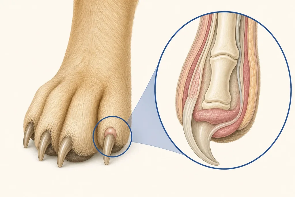
A nail-bed lesion can resemble infection, trauma, immune disease, or neoplasia. Persistence despite appropriate initial care, progressive swelling, nail loss, bleeding, or lameness raises the value of radiographs because bone lysis changes the differential and the scale of local treatment being considered. Radiographic change is still not a histologic diagnosis, and lack of obvious lysis does not make a growing soft-tissue lesion benign.
The diagnostic record should identify the exact digit and nail bed, duration, prior antimicrobial or anti-inflammatory exposure, regional lymph-node findings, and how the lesion affects weight bearing. Tissue collection is planned to preserve enough representative material while accounting for pain and function. When surgery is discussed, pathology of the removed tissue and an explicit margin assessment remain important; the visual appearance of the digit cannot substitute for classification.
Questions for the veterinary team
- Do radiographs show bone involvement?
- What sample best distinguishes infection from tumor?
- Should the draining node be sampled?
- Would amputation be diagnostic, therapeutic, or both?
- What wound and mobility plan follows?
Medical disclaimer: This article is educational and cannot diagnose, treat, or determine prognosis for an individual dog. It does not replace an examination or an individualized plan from a licensed veterinarian.