Canine Gastrointestinal Masses: Choosing Endoscopic, Image-Guided, or Surgical Tissue
Summary
Gastrointestinal tumors can share nonspecific signs and imaging features; lesion layer, location, obstruction, perforation risk, and suspected tumor class determine the most informative biopsy route.
Article
Evidence-informed clinical review. Vomiting, weight loss, diarrhea, anemia, reduced appetite, or an intestinal mass does not identify adenocarcinoma, lymphoma, gastrointestinal stromal tumor, smooth-muscle tumor, inflammation, or another process. The sampling route matters because superficial mucosal biopsies and full-thickness tissue answer different questions.
The clinical question
The team must identify where the lesion sits, whether it is focal or diffuse, which bowel layers are involved, whether regional nodes or other organs are abnormal, and whether obstruction, bleeding, or perforation changes urgency. The best specimen is the least burdensome one that can still answer the decision-driving question.
What current evidence can establish
Ultrasound, endoscopy, and CT provide different views of wall layers, lumen, extraintestinal structures, and surgical anatomy. Fine-needle samples may support lymphoma or sample nodes and liver, while histopathology is often needed to separate epithelial, lymphoid, and mesenchymal tumors. Immunohistochemistry or molecular testing may refine selected diagnoses.
A decision-focused pathway
Stabilize dehydration, pain, electrolyte disturbance, anemia, or obstruction before treating the case as an elective biopsy problem. Discuss whether endoscopic tissue can reach the lesion and include adequate depth. If surgery is likely regardless, plan resection, node or liver sampling, specimen orientation, and postoperative nutrition rather than collecting disconnected tests.
After diagnosis, distinguish local problems from systemic disease. Surgery may address a resectable nonlymphomatous mass or obstruction, while lymphoma generally prompts a systemic-treatment discussion. Supportive care for nausea, pain, intake, and stool losses belongs beside tumor-directed decisions and should have measurable targets.
Evidence gaps and interpretation
Imaging patterns overlap, a normal-appearing segment can still harbor disease, and small biopsies can miss deeper or heterogeneous lesions. Studies combine different sites and histologies, so a broad gastrointestinal-cancer statistic is rarely suitable for one dog. Nondiagnostic tissue is an information limit, not a benign result.
Safety, monitoring, and escalation
Repeated vomiting, abdominal distension, severe pain, black or bloody stool, collapse, or inability to keep water down requires prompt care. Biopsy and surgery can cause hemorrhage, leakage, peritonitis, aspiration, or delayed feeding. Exact fasting and medication directions must come from the procedural team.
Choose a specimen that reaches the abnormal layer
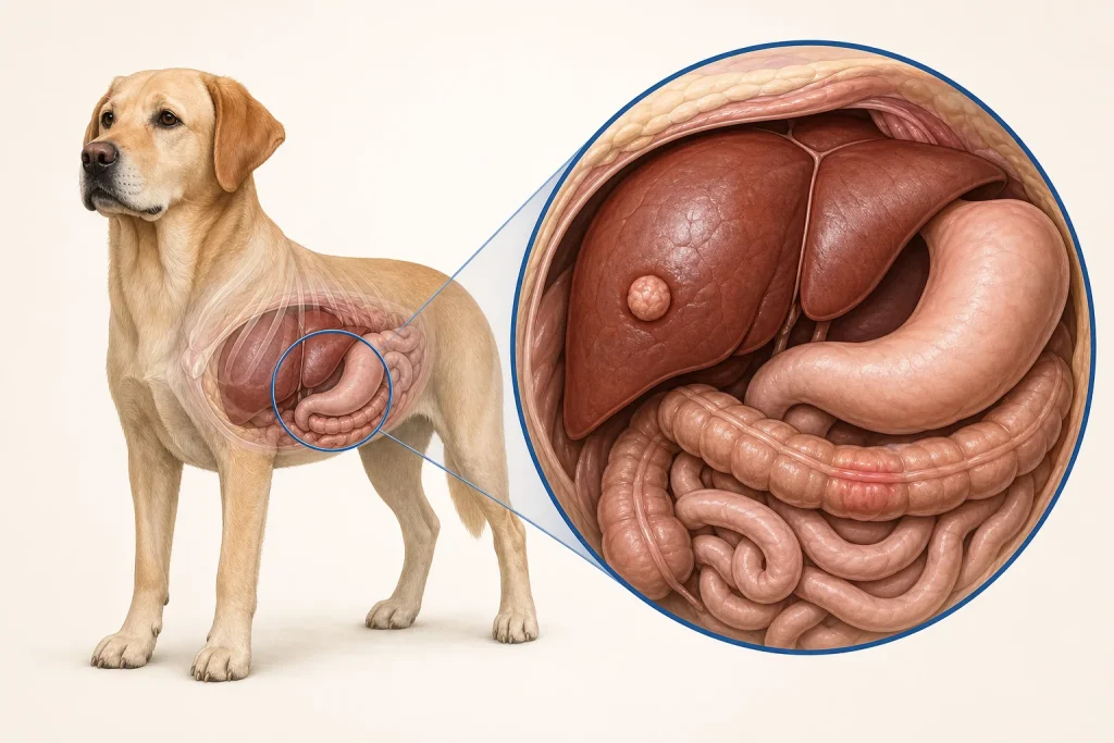
Gastrointestinal tumors may be mucosal, arise deeper in the bowel wall, or extend outside the lumen. That location matters because a visually abnormal surface can be sampled endoscopically, while a submucosal or outward-growing lesion may yield superficial material that misses the diagnostic tissue. Ultrasound or cross-sectional imaging can clarify wall layering, length of involvement, obstruction, regional nodes, and a possible percutaneous or surgical route.
The decision also includes the dog's stability, vomiting or bleeding, ability to pass food and fluid, risk of perforation, and whether exploration could be both diagnostic and therapeutic. A nondiagnostic biopsy should prompt review of depth, site, preservation, and pathology question rather than automatic repetition of the same method. Families benefit when the plan distinguishes obtaining any cells from obtaining architecture sufficient to classify the lesion and guide the next step.
Questions for the veterinary team
- Which bowel layer and location must the sample reach?
- Could endoscopy miss the suspected process?
- Is obstruction or perforation changing urgency?
- Which additional tissues should be sampled at surgery?
- What nutrition and complication plan follows the procedure?
Medical disclaimer: This article is educational and cannot diagnose, treat, or determine prognosis for an individual dog. It does not replace an examination or an individualized plan from a licensed veterinarian.