Canine Cutaneous Mast Cell Tumors: Why Appearance Does Not Predict Behavior
Summary
A structured owner review of mast cell tumor sampling, degranulation, grade, regional lymph nodes, surgical planning, and evidence boundaries.
Article
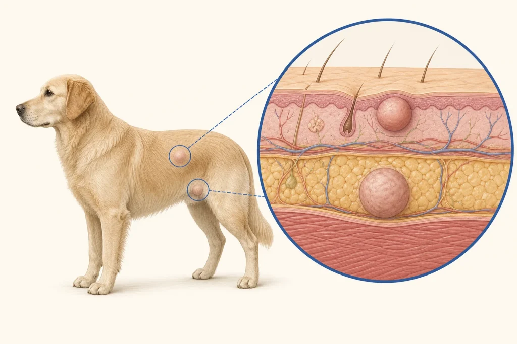
Structured owner review. Canine mast cell tumors illustrate why a photograph cannot diagnose or grade a lump. They can be small and movable, broad and inflamed, ulcerated, hairless, or hidden beneath the skin. Some fluctuate in size as mast-cell granules release inflammatory substances. Benign lesions and other tumors can mimic them, while an innocent-looking bump can still require treatment planning.
Recognition and initial sampling
Any new or changing mass deserves veterinary assessment. Fine-needle aspiration and cytology often identify mast cells because their granules have a characteristic microscopic appearance. Atypical or poorly granulated cases may require tissue and additional tests. Repeated squeezing or vigorous handling is unhelpful and may provoke local swelling, redness, or discomfort.
Inflammatory mediators released by mast cells can also contribute to vomiting, poor appetite, abdominal discomfort, or dark digested blood in stool in some patients. These signs require veterinary contact. Collapse, pale gums, breathing difficulty, severe weakness, or uncontrolled bleeding requires urgent care.
Why histopathology still matters
Cytology can often establish the tumor type, but histopathology evaluates tissue architecture and grade. Grade, mitotic features, invasion, location, size, ulceration, molecular findings when indicated, stage, and surgical margins may all contribute to risk assessment. No single feature should be converted into an exact outcome for an individual dog.
Staging should follow biology
Mast cell tumors commonly spread first to regional lymph nodes rather than the lungs. AAHA recommends selecting staging tests according to tumor behavior and whether results change management. A regional lymph node may contain metastatic cells even if it is not enlarged, and an enlarged node may be reactive rather than metastatic. Fine-needle sampling can therefore be more informative than size alone when nodal status matters.
Broader evaluation may be considered for large, rapidly growing, high-grade, recurrent, mucosal, clinically concerning, or node-positive tumors, or when systemic signs exist. The exact plan may include laboratory testing, lymph-node cytology, abdominal imaging, or targeted organ sampling. A small, slowly growing lesion may warrant a different plan. Owners should ask why each site is being checked.
Local treatment planning
Surgery is often used for local disease, with margins selected for tumor size, grade, location, and adjacent tissue. The visible edge does not necessarily represent the microscopic edge. The first planned excision offers the best chance to remove the tumor in an appropriate manner, so a casually shelled-out lump can reduce later options. The entire specimen should go to histopathology for confirmation, grade, and margin assessment.
When complete surgery is not feasible or pathology shows concerning features, the veterinary team may discuss scar re-excision, radiation, systemic therapy, local injectable treatment in eligible cases, monitoring, or palliative care. These are case-dependent choices, not interchangeable remedies.
Owner questions after diagnosis
- Was the diagnosis made by cytology, histopathology, or both?
- Which grading system was used, and what other prognostic features were reported?
- Which lymph node is regional to this mass, and was it sampled?
- Were surgical margins evaluated and oriented?
- What monitoring schedule fits this tumor's risk?
- What gastrointestinal, allergic-type, wound, or systemic signs require an urgent call?
Evidence boundary
Breed tendencies, grade categories, and group survival estimates cannot predict one dog's course. Treatment decisions should integrate the complete pathology report, stage, anatomical constraints, comorbidities, comfort, and family goals. Supplements or topical products should not replace sampling or oncologic planning.
Medical disclaimer: This article is general education and cannot diagnose, grade, stage, or treat a mast cell tumor. Do not manipulate a suspicious mass or give antihistamines, steroids, stomach medicines, or supplements without veterinary direction. Seek emergency care for collapse, breathing difficulty, pale gums, severe weakness, or uncontrolled bleeding.