Canine Nasal Tumors: Why CT, Biopsy, and Radiation Planning Answer Different Questions
Summary
A clinically cautious explanation of how anatomy, tissue diagnosis, staging, and radiation planning fit together in a dog with persistent nasal signs.
Article
Educational medical disclaimer: This article is for general learning only. Persistent nosebleeds, difficult breathing, facial swelling, neurologic change, or rapid decline require veterinary assessment; the text cannot diagnose an individual dog.
Chronic nasal discharge, sneezing, noisy airflow, or blood from one nostril can arise from cancer, fungal disease, inflammation, a foreign body, dental disease, or other causes. Because several conditions look alike at home, a nasal-tumor pathway should not begin with an assumption. It begins by defining urgency, mapping the anatomy, and obtaining representative tissue when it is safe and useful.
The examination frames the problem
The veterinarian will consider the duration and side of the signs, airflow, facial symmetry, oral and dental findings, eye changes, regional lymph nodes, pain, and the dog's general health. Bloodwork may help assess anesthesia risk or concurrent disease but does not identify most nasal masses. Severe bleeding, open-mouth breathing, blue or gray mucous membranes, collapse, or acute neurologic signs warrant emergency care.
CT maps structure
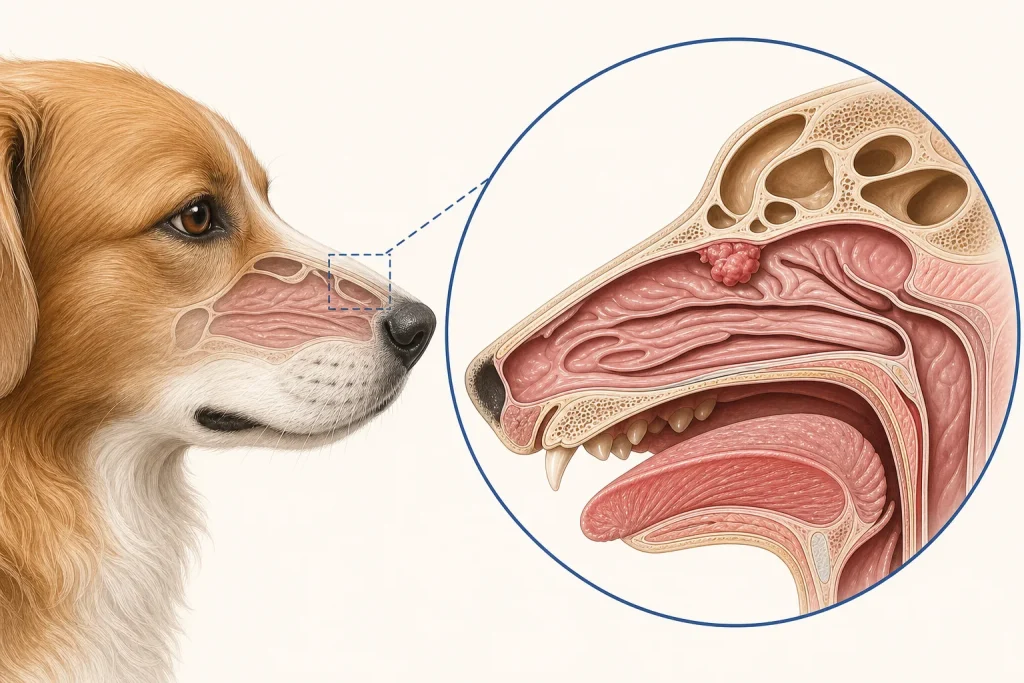
Computed tomography can show which nasal passages and sinuses are involved, whether bone has been destroyed, and how close the process lies to the orbit, palate, or skull. Contrast images may add information about tissue and vessels. These details can guide rhinoscopy, biopsy, staging discussions, and radiation planning. Yet CT appearance alone may not distinguish a malignant tumor from fungal or severe inflammatory disease in every dog. The correct language is often compatible with or suspicious for, not proven by imaging.
Biopsy names the process
Rhinoscopy, image-guided sampling, or another biopsy technique may be selected according to location and safety. Nasal tissue can be fragile and vascular, so bleeding risk, airway management, and recovery planning matter. Superficial inflammation or necrosis may surround a deeper lesion; a nondiagnostic sample may therefore require pathology review, additional stains, cultures, or repeat targeted sampling. Families should ask whether the specimen is representative and whether the report gives a firm diagnosis or a differential list.
Staging and treatment planning are separate jobs
Once a tumor type is established, the team can choose staging tests based on its expected behavior and the family's goals. Regional lymph-node assessment and thoracic imaging may be discussed. A normal-sized node is not guaranteed to be free of tumor, while an enlarged one may be reactive. Conversely, no visible distant disease on selected tests cannot exclude microscopic spread.
Radiation planning uses CT data in a different way: clinicians define a treatment target and nearby normal structures, then design dose delivery. Studies of conventionally fractionated and stereotactic approaches report outcomes in groups of dogs, but they do not establish that one schedule is best for every diagnosis or anatomy. Protocol choice depends on pathology, extent, goals, equipment, expected effects, anesthesia burden, and clinician judgment. Surgery and systemic therapy have roles in selected cases, while symptom-focused care may be appropriate when definitive local treatment does not fit.
Ask about effects across time
Radiation discussions should separate effects that may appear during or soon after treatment from later effects. The eyes, skin, oral tissues, brain, and other nearby structures may influence the plan. Owners need written instructions for expected signs, medications, feeding support, emergency thresholds, and follow-up. A statement such as radiation is targeted should not be heard as risk-free.
Bring these questions
- Which diagnoses can still mimic the imaging pattern?
- Where will tissue be collected, and how will bleeding and airway risks be managed?
- What did staging evaluate, and which result would alter the plan?
- Is the aim durable local control, symptom relief, or both?
- How many anesthetic events are expected, and what monitoring is used?
- What changes at home should trigger an urgent call?
A useful plan connects every test to a decision and every treatment to an observable goal such as easier airflow, reduced bleeding, preserved appetite, or sustained comfortable activity. Follow-up should track both tumor-related signs and treatment effects rather than relying on a single scan.