Canine Melanocytic Tumors: Why Oral, Digital, and Haired-Skin Sites Are Not Interchangeable
Summary
A careful review of how anatomic site, histopathology, proliferation measures, and staging combine when a dog has a melanocytic tumor.
Article
Educational medical disclaimer: This article offers general education only. It cannot classify a pigmented lesion, interpret a pathology report, or recommend treatment for an individual dog; those tasks require a licensed veterinarian and appropriate tissue evaluation.
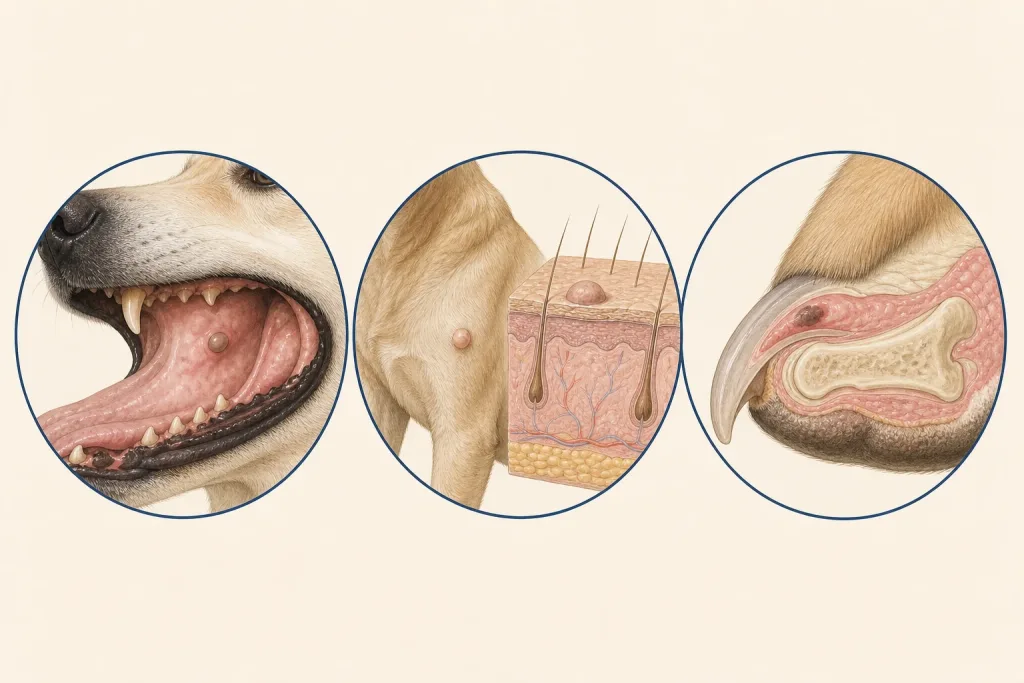
The word melanoma can make every dark spot feel dangerous, yet color alone does not establish that a lesion is melanocytic, and melanocytic tumors do not behave identically across the body. Tumors of the oral cavity, nail unit or digit, and haired skin arise in different clinical settings. Anatomic site is important, but it is not a perfect substitute for pathology, stage, and the dog's actual course.
Begin with accurate localization
The record should specify where the lesion starts, not merely where it is visible. An oral mass may involve gingiva, lip, palate, tongue, or another structure. A digital lesion may arise from skin, nail bed, or deeper tissue. A haired-skin nodule can be dermal or subcutaneous. Size, ulceration, fixation, pain, bone involvement, and regional nodes should be documented before treatment. Photography and imaging can support this map, but neither names the cell type.
Pigment is an unreliable shortcut
Some melanocytic tumors contain obvious dark pigment; others are amelanotic and resemble unrelated tumors. Conversely, several benign or inflammatory lesions are pigmented. Cytology can be helpful in some cases, but histopathology is generally needed to evaluate architecture and features associated with behavior. Pathologists may use immunohistochemistry when routine appearance does not clearly establish melanocytic origin.
Pathology adds more than a label
The international veterinary oncology consensus on canine and feline melanoma and the pathology consensus for canine melanocytic neoplasms emphasize integrating site with microscopic features. Depending on specimen and laboratory approach, a report may address mitotic activity, cellular atypia, invasion, ulceration, pigmentation, lymphatic involvement, and margins. Some laboratories may report additional proliferation markers when validated and clinically relevant. Owners should ask which measurements were made, whether the specimen was complete enough to support them, and how strongly they apply at that anatomic site.
No single feature should be translated into an exact countdown. Site is a major prognostic variable, but individual behavior varies. A small lesion at a higher-risk site is not automatically harmless, and a concerning microscopic feature does not prove that spread is already present.
Choose staging that fits the diagnosis
Regional lymph nodes deserve attention even when they are not enlarged. Sampling may be more informative than size alone when nodal status changes the plan. Thoracic imaging and other anatomic imaging may be considered based on the primary site, pathology, clinical findings, and family goals. For oral or digital disease, CT or radiographs can help define bone involvement and plan local treatment. No-evidence-of-disease on selected tests means disease was not detected at their resolution; it does not rule out microscopic cells.
Local and systemic strategies have different aims
Surgery seeks local control and may provide the definitive specimen. The margin needed and likely functional effect depend on site and anatomy. Radiation may be considered when complete surgery is not feasible, after selected incomplete excisions, or for palliation. Systemic approaches may be discussed in cases with higher metastatic risk or documented spread. Evidence strength, availability, cost, visit burden, and uncertainty should be stated explicitly. A therapy being biologically plausible or commercially available does not prove benefit for every tumor.
Questions that improve the report discussion
- Is melanocytic origin confirmed, and was immunohistochemistry required?
- What is the exact primary site and depth?
- Which microscopic features are validated for prognosis at this site?
- Were margins assessable, and how should they influence local-control choices?
- Which regional node drains this area, and was it sampled?
- What is known about distant disease, and what remains below detection?
- How will local function, pain, eating, walking, and quality of life be followed?
A coordinated plan preserves uncertainty where it truly exists. It avoids assuming that all dark lesions are melanoma, that all cutaneous melanocytic tumors are harmless, or that site alone decides outcome. New oral bleeding, difficulty eating or breathing, rapidly worsening lameness, uncontrolled pain, or sudden decline should prompt urgent veterinary contact.