A Solitary Pulmonary Mass in a Dog: Primary Tumor, Metastasis, or Another Disease?
Summary
A lung mass on imaging is an anatomic finding, not a histologic diagnosis; origin, regional lymph nodes, other lesions, respiratory stability, and tissue feasibility shape the next decision.
Article
Evidence-informed clinical review. A solitary lung lesion can represent a primary pulmonary tumor, metastasis from a cancer elsewhere, or a non-neoplastic process. The label matters because staging, surgical candidacy, prognosis, and systemic-treatment discussions differ. Imaging can organize those possibilities, but it usually cannot name the tissue with certainty.
The clinical question
The practical question is not simply whether a nodule looks malignant. The team needs to ask whether it is solitary or part of a wider pattern, whether thoracic lymph nodes or pleura are involved, whether another primary tumor is known, and whether the dog can safely undergo sampling or lung-lobe surgery.
What current evidence can establish
Thoracic radiographs may identify a lesion, while CT can better define number, location, airway or vessel relationships, lymph nodes, and other small pulmonary abnormalities. Definitive classification generally depends on cytology or histopathology. Even then, a pathologist may need the dog’s prior tumor history and immunohistochemistry to assess likely origin.
A decision-focused pathway
Begin with respiratory assessment and review of every previous pathology report. Compare current imaging with earlier studies when available. If local treatment is being considered, obtain imaging that answers resectability and node questions, and discuss whether a node should be sampled. Choose a tissue route only after balancing diagnostic yield against pneumothorax, hemorrhage, anesthesia, and lesion accessibility.
Before surgery, define what would make the operation diagnostic, potentially controlling, or inappropriate. After resection, the report should address tumor type, grade where meaningful, margins, vascular or lymphatic invasion, and node findings if sampled. Those details, not the phrase lung cancer alone, guide surveillance and any systemic-therapy consultation.
Evidence gaps and interpretation
Published outcomes combine different histologies, stages, imaging eras, and referral populations. A single apparently isolated mass does not prove an early primary tumor, and multiple nodules do not automatically identify the source. Conversely, a negative radiograph cannot exclude microscopic pulmonary disease. The confidence of every inference should be made explicit.
Safety, monitoring, and escalation
Breathing difficulty, blue or gray mucous membranes, collapse, coughing blood, or rapidly worsening effort requires urgent stabilization. Sampling and thoracic surgery carry patient-specific respiratory, bleeding, pain, and anesthesia risks. Written postoperative and pathology follow-up should be in place before discharge rather than added after a complication.
Read the thoracic pattern before naming the mass
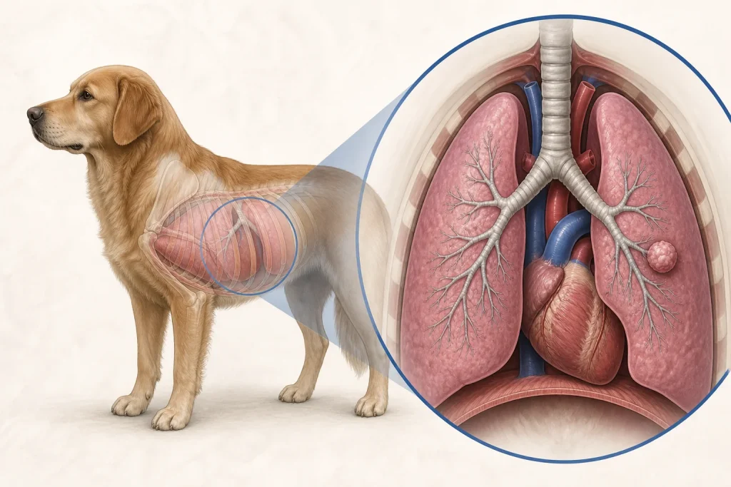
A solitary pulmonary mass is a radiographic description, not a tissue diagnosis. Interpretation changes when the study also shows additional nodules, enlarged intrathoracic lymph nodes, pleural fluid, airway involvement, or continuity with the chest wall. The dog's prior tumor history matters, but it cannot by itself prove that a new lung lesion is metastatic. A complete thoracic review helps keep primary lung cancer, metastasis, inflammatory disease, and other causes open until stronger evidence separates them.
Imaging is most useful when it answers a defined next question: whether there is a safe sampling route, whether apparently localized surgery is technically plausible, and whether disease is present elsewhere. Cytology or histopathology may still be needed to establish identity. If a sample is nondiagnostic, the team can revisit lesion location, sample quality, procedural risk, and whether the result would alter treatment rather than treating an uncertain label as final.
Questions for the veterinary team
- What evidence favors a primary lung lesion versus metastasis or inflammation?
- Would CT or node sampling change surgical planning?
- What tissue method offers useful yield at acceptable risk?
- Which pathology features will drive the next decision?
- What respiratory changes require emergency care?
Medical disclaimer: This article is educational and cannot diagnose, treat, or determine prognosis for an individual dog. It does not replace an examination or an individualized plan from a licensed veterinarian.