A Canine Renal Mass: Characterize Both Kidneys Before Planning Nephrectomy
Summary
A renal mass may be primary, metastatic, multicentric, inflammatory, or cystic; both-kidney function, vascular anatomy, metastatic assessment, and tissue identity determine whether removal is reasonable.
Article
Evidence-informed clinical review. Kidney tumors are uncommon in dogs, and a mass may be found during imaging for unrelated signs. A unilateral lesion, bilateral nodules, and diffuse enlargement suggest different possibilities, but enhancement pattern alone cannot supply a final diagnosis. Any plan that may remove one kidney must first understand the other kidney and the whole patient.
The clinical question
The team needs to determine whether the lesion is truly renal, whether one or both kidneys are involved, whether vessels or adjacent organs are affected, and whether disease exists elsewhere. The next decision is whether tissue should be obtained before surgery or whether nephrectomy would provide both diagnosis and local treatment.
What current evidence can establish
Urinalysis, urine culture where indicated, blood pressure, CBC, chemistry, and urine protein assessment describe function and complications but rarely name a mass. Ultrasound and CT define laterality, distribution, enhancement, vessels, nodes, and metastasis. Cytology may identify selected diseases such as lymphoma; histopathology often supplies definitive classification.
A decision-focused pathway
Review prior creatinine, urine concentration, protein loss, blood pressure, infections, blood in urine, pain, and imaging. Assess the contralateral kidney rather than assuming it is normal. If percutaneous sampling is considered, define hemorrhage risk, expected yield, route, and whether the result would avoid or alter surgery.
For a potentially resectable unilateral primary tumor, surgical consultation should address vascular anatomy, metastatic findings, residual renal reserve, perioperative monitoring, and pathology handling. Bilateral or multicentric disease may shift attention toward systemic diagnosis and therapy. Comfort, appetite, hydration, and urinary function remain relevant across every path.
Evidence gaps and interpretation
Imaging features overlap among carcinoma, lymphoma, sarcoma, metastasis, benign lesions, and inflammatory disease. Recent CT research can describe associations, not deliver certainty for one scan. Retrospective surgical studies select cases and should not be read as a guarantee that nephrectomy is appropriate or beneficial for every renal mass.
Safety, monitoring, and escalation
Inability to urinate, collapse, severe abdominal pain, repeated vomiting, marked weakness, or visible heavy bleeding in urine needs prompt assessment. Biopsy and surgery can cause hemorrhage, urine leakage, kidney injury, hypertension, pain, and anesthesia complications. Human pain medicines can worsen kidney risk.
The opposite kidney is part of every local-treatment decision
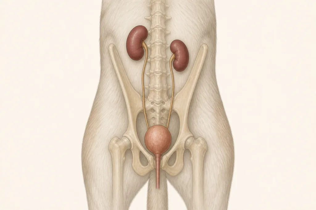
A renal mass assessment is incomplete if it describes only the affected side. Imaging should characterize both kidneys, renal vessels, ureters, regional nodes, abdominal spread, and any venous extension. Laboratory and urine findings add information about overall renal function and concurrent urinary disease, while neither imaging nor laboratory data alone establish the exact histologic type.
Before nephrectomy is considered, the record should connect contralateral structure and function, anesthetic risk, suspected distribution, and expected diagnostic value of the removed tissue. Bilateral or multifocal findings create a different problem from a solitary resectable lesion. If biopsy is proposed instead, the team can explain the bleeding risk, route, specimen requirement, and which result would redirect care. Post-procedure monitoring then needs a renal-function baseline that can be compared over time.
Questions for the veterinary team
- Are both kidneys structurally and functionally assessed?
- What diagnoses remain plausible from imaging?
- Would preoperative tissue change the plan?
- Does CT show vascular or metastatic disease?
- How will renal function and blood pressure be monitored afterward?
Medical disclaimer: This article is educational and cannot diagnose, treat, or determine prognosis for an individual dog. It does not replace an examination or an individualized plan from a licensed veterinarian.