Suspected Appendicular Osteosarcoma: From Painful Limb to Tissue, Stage, and Local Control
Summary
A stepwise review of how clinicians approach a destructive long-bone lesion without treating an imaging impression as a final diagnosis.
Article
Educational medical disclaimer: This review is general education, not a diagnosis or treatment plan. A painful limb, sudden inability to bear weight, or suspected fracture requires prompt examination by a licensed veterinarian.
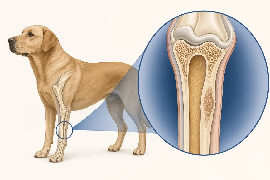
When radiographs show an aggressive lesion in a long bone, osteosarcoma may be high on the differential list, but an image describes a pattern rather than naming every possible cause. Infection, another primary bone tumor, metastatic disease, and less common disorders can resemble one another. A careful pathway separates four questions: Is the bone structurally safe today? What is the lesion? Is detectable disease present elsewhere? Which local-control and systemic-care options fit this dog and family?
First protect the dog and the bone
Pain control and safe movement come before a complete oncology workup. A weakened bone can fracture with ordinary activity. Until the veterinarian defines risk, families may be asked to restrict running and jumping, use secure footing, and assist with a harness. This is individualized advice; an owner should not splint a limb or change medication without instructions. Sudden severe pain, a new limb angle, collapse, pale gums, or distress is an emergency.
Imaging establishes suspicion, not certainty
Initial radiographs usually include the affected region and may show bone loss, new irregular bone, cortical disruption, or soft-tissue change. The American College of Veterinary Surgeons notes that bone-tumor evaluation can also involve chest imaging and other tests selected for the case. More advanced imaging may help define anatomy, evaluate a complex site, or plan a procedure. None of these automatically supplies histologic identity.
A veterinarian may recommend a fine-needle sample or bone biopsy. Sampling must be planned to avoid a major vessel or nerve, reduce fracture risk, and place the tract where it can be removed during later definitive surgery if necessary. Results should be interpreted alongside imaging and clinical findings. If infection is plausible, culture or additional testing may be important. An inconclusive sample is a limitation of that sample, not proof that the lesion is benign.
Stage with a purpose
Staging looks for clinically detectable spread and establishes a baseline. The exact package depends on the lesion, the dog's health, and what the family might choose. Thoracic imaging is commonly discussed because the lungs are a relevant site for many bone tumors. Clinicians may assess regional nodes, laboratory values, or other painful skeletal sites when findings suggest a reason. No-evidence-of-metastasis means that the selected tests did not detect spread; it cannot exclude microscopic disease.
Separate local control from systemic risk
Local control addresses the painful primary lesion. Limb amputation is one established option for appropriately selected patients, and ACVS describes candidacy as a whole-dog assessment rather than an age test. Limb-sparing procedures or radiation approaches may be considered in selected cases depending on location, anatomy, expertise, goals, and expected complications. Systemic therapy addresses risk beyond the primary site. The AAHA oncology guidance emphasizes matching modalities to tumor biology, patient condition, evidence, and family priorities.
Families deserve a balanced discussion of function, pain, visit frequency, adverse effects, rehabilitation, cost, and what will happen if the first plan does not work. Videos of the dog moving at home, an orthopedic and neurologic assessment of the other limbs, body condition, and concurrent disease can make that discussion more concrete.
A decision-ready consultation
- What findings make this lesion suspicious, and what alternatives remain?
- Does the result need tissue confirmation before definitive treatment in this case?
- What is the current fracture risk, and what activity is safe?
- Which staging result could change the local or systemic plan?
- What are realistic best-, expected-, and worst-case functional outcomes?
- How will pain and quality of life be measured at home?
The most responsible plan may be multimodal treatment, a local palliative approach, or comfort-focused care. Choosing among them is not a test of devotion. It is a shared decision built around the dog's comfort, the evidence that applies, and the family's practical limits.