Canine Oral Masses: A Function-Preserving Diagnostic and Planning Pathway
Summary
An evidence-informed pathway for oral masses that connects biopsy and imaging with eating, pain, airway safety, surgical planning, and owner goals.
Article
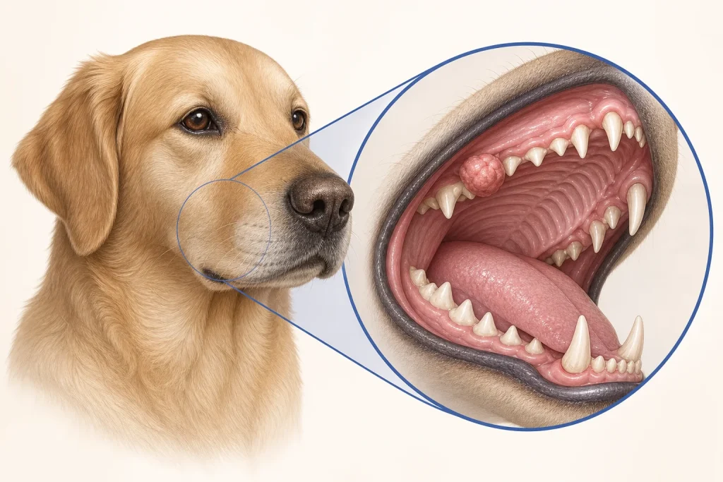
Evidence-informed clinical review for owners. An oral mass is not a diagnosis. Growths can arise from gums, bone, dental tissues, salivary tissues, cheek, palate, tongue, or tonsil, and benign and malignant lesions may look alike. Because the mouth is essential for eating, drinking, breathing, grooming, and social behavior, diagnosis and treatment planning must consider function from the beginning.
Clinical presentation
Owners may notice a visible growth, bad breath that persists despite dental care, drooling, blood on toys or in the water bowl, dropping food, chewing on one side, difficulty swallowing, facial asymmetry, loose teeth, reduced appetite, or weight loss. Sneezing, nasal discharge, or bleeding can occur when a lesion involves nearby structures. These signs also occur with dental disease, trauma, or infection. Their presence justifies an examination; it does not identify cancer.
Examination and stabilization
The veterinarian assesses the oral cavity as safely as the dog permits, along with facial symmetry, regional lymph nodes, pain, hydration, body condition, and airway. A complete examination may require sedation or anesthesia because important areas lie beneath the tongue or toward the throat. Active hemorrhage, breathing difficulty, inability to swallow, severe pain, or rapid decline changes the priority to urgent stabilization.
Obtaining a diagnosis
Cytology may help in selected lesions or lymph nodes, but tissue biopsy is commonly needed to determine the oral tumor type. The biopsy should be positioned so its tract can be removed or incorporated into later treatment when necessary. Surface appearance can underestimate deeper invasion: Cornell describes an iceberg effect for oral squamous cell carcinoma, in which tissue and bone involvement may extend beyond what is visible.
Histopathology may identify tumor type, grade when applicable, invasion, and other prognostic features. An inconclusive sample should prompt a discussion of whether it was representative, whether additional tissue or special stains are needed, and whether review by a veterinary pathologist would change the plan.
Imaging and staging
Dental radiographs can reveal bone involvement near teeth. CT may better define three-dimensional extent for surgery or radiation planning. Depending on diagnosis, staging may include lymph-node sampling, chest imaging, laboratory tests, or other targeted evaluation. Node size alone cannot reliably confirm or exclude metastasis. Each proposed test should be connected to a decision about feasibility, prognosis, or treatment.
Treatment planning through function
Surgery may range from soft-tissue excision to removal of part of the mandible or maxilla. Radiation, systemic therapy, or comfort-focused care may be considered depending on histology, location, stage, margins, and family goals. Ask how the option may affect eating, drinking, tongue movement, jaw strength, appearance, pain, anesthesia exposure, and home care. Many dogs can adapt after substantial oral surgery, but outcomes are procedure- and patient-specific and must not be promised.
Decision questions
- Where exactly does the lesion arise, and is bone involved?
- What sample will provide a reliable diagnosis without compromising later treatment?
- Which lymph nodes drain this location, and should they be sampled?
- What local-control options are anatomically possible?
- What functional change and recovery burden should we expect?
- How will pain, nutrition, and oral hygiene be supported?
Evidence limits
Oral cancer is not one disease. Statistics for one histologic type or location should not be applied to another. Even after imaging, microscopic extent may differ from visible extent. Prognosis should integrate pathology, anatomical site, stage, local-control feasibility, and the dog's overall condition.
Medical disclaimer: This article is educational and cannot diagnose an oral lesion or recommend biopsy, imaging, surgery, radiation, or medication for an individual dog. Seek prompt veterinary examination for any oral mass, unexplained bleeding, eating difficulty, or facial swelling; breathing difficulty or uncontrolled bleeding is an emergency.