Canine Orbital Masses: Protect Vision While Defining Extent and Tissue
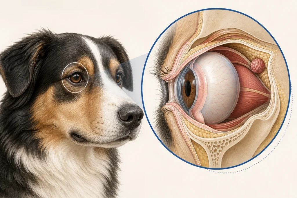
Summary
Forward displacement of an eye, eyelid swelling, or altered globe movement can arise from tumor, inflammation, hemorrhage, or adjacent disease; protecting the cornea and defining anatomy are simultaneous priorities.
Article
Evidence-informed clinical review. The orbit is a confined space shared by the globe, muscles, nerves, vessels, lacrimal tissue, bone, sinuses, nasal structures, and nearby teeth. A growing lesion can impair blinking or vision before its origin is clear. Acute painful disease and slower neoplasia can overlap, so examination, imaging, and tissue must be integrated.
The clinical question
The first questions are whether vision or the ocular surface is threatened, whether infection or hemorrhage requires urgent management, and which adjacent compartments are involved. The longer-term question is whether local treatment can preserve comfort and function and what tissue diagnosis is needed before committing to surgery or radiation.
What current evidence can establish
A complete ophthalmic and oral examination can localize clues but cannot see the full orbit. Ultrasound may assess accessible structures; CT is useful for bone, sinus, and surgical anatomy; MRI may add soft-tissue and neurologic detail. Histopathology defines tumor type, while systemic staging is tailored to that diagnosis.
A decision-focused pathway
Document onset, pain opening the mouth, nasal signs, dental history, vision change, globe position, and exposure injury. Protect an incompletely closing eye under veterinary direction while diagnostics proceed. The ophthalmologist, radiologist, surgeon, and oncologist should agree on a sampling route that preserves later options and avoids blind passage through critical structures.
Local choices may include limited excision, removal of the eye, more extensive orbital surgery, radiation, systemic treatment, or comfort care depending on origin and extent. Appearance after surgery, likelihood of vision preservation, pain, anesthesia burden, and need for repeated visits deserve direct discussion before consent.
Evidence gaps and interpretation
Orbital masses are heterogeneous and relatively uncommon. Imaging may suggest a compartment or tissue of origin but cannot guarantee histology. Published outcome estimates can overrepresent referral cases and specific procedures. Preserving the globe is not synonymous with preserving useful, comfortable vision, and removal is not automatically definitive cancer control.
Safety, monitoring, and escalation
Sudden eye protrusion, severe ocular pain, inability to close the eyelids, acute vision loss, bleeding, neurologic change, or systemic illness needs urgent assessment. Topical human products and pressure on the globe can worsen injury. Diagnostic and treatment procedures carry vision, hemorrhage, nerve, infection, and anesthesia risks.
Protect function while defining the orbital compartment
Orbital disease can present as globe displacement, pain on opening the mouth, eyelid or conjunctival swelling, reduced vision, or neurologic change. Recording which functions are affected helps establish urgency and provides a baseline that a size measurement alone cannot. Examination may suggest whether disease is within the orbit, arises from adjacent nasal or oral structures, or involves bone, but deep boundaries generally require advanced imaging.
The imaging question should include the optic pathway, extraocular muscles, orbital bones, nasal cavity, skull, and a safe route to representative tissue. Sampling plans must account for the eye and nearby vessels and nerves. If preservation of vision is a goal, that goal needs to be stated before a procedure is chosen. A coordinated ophthalmic, imaging, surgical, and pathology interpretation reduces the risk of treating a location label as though it were already a tumor diagnosis.
Questions for the veterinary team
- Is the cornea or vision in immediate danger?
- Which orbital compartment appears involved?
- Would CT, MRI, or both change the procedure?
- How will tissue be obtained without compromising local options?
- What comfort and appearance changes should the family expect?
Medical disclaimer: This article is educational and cannot diagnose, treat, or determine prognosis for an individual dog. It does not replace an examination or an individualized plan from a licensed veterinarian.