Canine Cardiac Masses and Pericardial Effusion: Stabilize Before Naming the Tumor
Summary
Pericardial fluid can impair cardiac filling and cause collapse; echocardiographic location informs the differential, but stabilization and diagnostic certainty are separate tasks.
Article
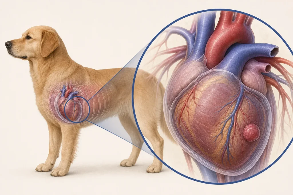
Evidence-informed clinical review. A dog with weakness, collapse, abdominal fluid, rapid breathing, or muffled heart sounds may have pericardial effusion with cardiac tamponade. Neoplasia is one important cause, but idiopathic and other diseases occur. Even when ultrasound shows a mass, location suggests possibilities rather than guaranteeing histology.
The clinical question
The first question is whether fluid pressure is compromising circulation and requires urgent drainage. After stabilization, the team can assess mass location, recurrence, other thoracic or abdominal disease, and whether additional sampling or imaging will change treatment. Emergency decisions should not wait for a perfectly named tumor.
What current evidence can establish
Echocardiography is central for detecting pericardial effusion, tamponade physiology, and some cardiac masses. Thoracic radiographs, abdominal imaging, CT, fluid analysis, and laboratory testing can add context. Pericardial-fluid cytology often has limitations, and a nondiagnostic sample cannot rule out neoplasia. Tissue confirmation may be difficult or unsafe.
A decision-focused pathway
Document episode timing, gum color, breathing, abdominal enlargement, exercise tolerance, and prior fainting. During acute care, stabilization and pericardiocentesis may precede staging. Once stable, identify whether the mass is right atrial, heart-base, or elsewhere; review recurrent effusion; and discuss cardiology, surgery, oncology, or palliative consultation.
Options may include repeated drainage, pericardial procedures, surgery in selected cases, systemic or targeted treatment, and comfort-focused care. Each option should have a stated goal: relieve tamponade, obtain tissue, reduce recurrence burden, control a defined tumor, or support quality of life. Access and expertise materially affect feasibility.
Evidence gaps and interpretation
Hemangiosarcoma and heart-base tumors are common neoplastic considerations in dogs, but imaging location is not infallible. Outcome reports mix tumor types and interventions and can be influenced by which dogs survive to referral or surgery. A negative fluid sample and a temporarily improved dog do not establish a benign cause.
Safety, monitoring, and escalation
Collapse, severe weakness, gray or pale gums, labored breathing, or a rapidly enlarging abdomen requires emergency care. Home activity and medication changes should follow the cardiology team’s directions. Invasive sampling, drainage, anesthesia, and surgery carry arrhythmia, bleeding, injury, recurrence, and hemodynamic risks.
Effusion physiology comes before tumor naming
When pericardial fluid impairs cardiac filling, weakness, collapse, rapid breathing, poor perfusion, or abdominal distension can reflect a time-sensitive hemodynamic problem. Stabilization and echocardiographic assessment take priority over completing an ideal tumor workup. The location of a visible mass, the amount and recurrence of fluid, cardiac function, and findings elsewhere help shape the differential, but location is not identical to histology.
Tissue sampling around the heart may carry substantial risk and may not always be the first or most useful step. The team can state what additional imaging, fluid analysis, or follow-up would change management and what uncertainty will remain without tissue. A recurrence plan should identify the signs that require immediate reassessment and how serial echocardiographic findings will be compared, rather than relying on one fluid measurement in isolation.
Questions for the veterinary team
- Is tamponade present and does the dog need urgent drainage?
- Where is the mass and how certain is that localization?
- What can fluid analysis answer here?
- Would tissue change a realistic treatment choice?
- What is the plan if effusion returns?
Medical disclaimer: This article is educational and cannot diagnose, treat, or determine prognosis for an individual dog. It does not replace an examination or an individualized plan from a licensed veterinarian.